What is spasticity
Spasticity is a motor disorder characterized by muscle hyperactivity (or hypertony). It produces involuntary contraction of the involved muscles.
Definition
Lance's Lance JW. Symposium synopsis. In: Feldman RG, Young RR, Koella WP, eds. Spasticity: disordered motor control. Chicago: Yearbook Medical 1980: 485-94. widely accepted definition of 1980 states: "Spasticity is a motor disorder characterized by a velocity-dependent increase in tonic stretch reflexes The stretch reflex (“myotatic reflex”) is an automatic (or “reflex”) muscle contraction in response to an unexpected stretching within the muscle. It is sought for during every neurological examination. The most known reflex is the patellar reflex (“knee-jerk): with the knee flexed and the thigh muscles relaxed, percussion of the patellar ligament causes leg extension by reflex contraction of the quadriceps muscle.
Stretch reflexes are exagerated in brain and spine lesions, especially in spasticity.(“muscle tone”) with exaggerated tendon jerks, resulting from hyper-excitability of the stretch reflex, as one component of the upper motoneuron* syndrome."
The SPASM Consortium (2006) produced a simpler definition:"Assuming that all involuntary activity involves reflexes, spasticity is an intermittent or sustained involuntary hyperactivity of a skeletal muscle associated with an upper motoneuron* lesion."
Spasticity is elective.
It involves only certain muscles.
In the upper limb, these are usually the adductor muscles (bringing the arm alongside the trunk), the flexor muscles (flexion of the elbow, the wrist and the fingers), and the pronator muscles (bringing the palm of the hand face down).
In the lower limb, it affects mainly the adductor muscles (bringing the legs close to each other), the flexor muscles of the hip and knee, and the muscles which extend the foot and rotate it inwards.
Non-spastic muscles are usually weak or paralysed.
Spasticity is elastic.
The more one tries to fight it, the more it increases. But it finally gives way, and the joint can be moved all the way, unless there is some muscle (or joint) contracture associated.
The stretch reflex is increased in spastic muscles, and can produce a clonusInappropriate recruitment of a muscle upon activating the muscle producing the opposite movement (antagonist). (example: co-contraction of the triceps when activating the biceps prevents flexion of the elbow).A clonus is a series of involuntary rhythmic muscle contractions. Unlike abnormal movements, it does not occur spontaneously, but is initiated by a reflex..
Spasticity is variable.
It increases with cold temperatures, emotions, fatigue. It is often milder at rest, and can disappear during sleep, if there is no associated muscle contracture.
How do we measure it?
Measurement of spasticity is essential to assess the response to treatment. But it is difficult to measure because of its multifactorial nature. Different methods are available for measurement but none of them is precise and reliable enough to quantify the severity of spasticity clinically.
- Ashworth scale (1964), modified by Bohannon and Smith Bohannon RW, Smith MB. (1987)
Interrater reliability of a modified Ashworth scale of muscle spasticity. Phys Ther 67:206-7. (1987) is validated only for the lower limb.
|
Score |
Modified Ashworth scale (Bohannon and Smith 1987) |
|
|
0 |
|
No increase in tone |
|
1 |
|
Slight increase in tone giving a catch, release and minimal resistance at the end of range of motion (ROM) when the limb is moved in flexion/extension |
|
1+ |
|
Slight increase in tone giving a catch, release and minimal resistance throughout the remainder (less than half) of ROM |
|
2 |
|
More marked increased in tone through most of the ROM, but limb is easily moved |
|
3 |
|
Considerable increase in tone – passive movement difficult |
|
4 |
|
Limb rigid in flexion and extension |
- The Tardieu scale a. Tardieu G, Shentoub S, Delarue R. (1954)
A la recherche d’une technique de mesure de la spasticité. Revue Neurol 91:143-4.
b. Gracies JM, Burke K, et al (2010)
Reliability of the Tardieu Scale for Assessing Spasticity in Children With Cerebral Palsy. Arch Phys Med Rehab. 91, 3: 421-428 measures the maximal angle obtained by mobilization of the joint by the examiner during as slow a movement as possible (V1), under gravitational pull (V2) and at a fast rate (V3: angle at which the examiner feels a “catch” in the muscle).
Spasticity may be associated with muscle / joint contractures, and with abnormal movements.
What is muscle contracture?
It is a consequence of spasticity. The muscle itself and its envelope (fascia) become permanently contracted, or tight. It becomes impossible to extend the involved joint fully.
Physiotherapy and splinting are important to prevent muscle contracture. But when it is established, it may require surgery.
What is an abnormal movement?
Not all spastic patients are affected by abnormal movements
Abnormal movements occur spontaneously and are not controlled by the patient . They can occur either when the patient is at rest (chorea, athetosis), or during voluntary movement (dyskinesia, dystonia) .
Distonia
Is sensation affected?
Sensation is affected in most, but not all cases. In cerebral palsy, the sensation of touch is generally unaffected, but complex sensations (sense of the position of the fingers [proprioception], recognition of an object placed in the hand [stereognosis]…) are often altered.
Who is affected by spasticity
Spasticity typically occurs in patients following cerebral palsy, stroke, brain injury (trauma and other causes, e.g. anoxia, post-neurosurgery), spinal cord injury, multiple sclerosis and other neurological diseases.
- Cerebral palsy is a brain damage which occurs either before birth, or during infancy (up to the age of 2 y.o.). It may be due to varied causes: temporary lack of blood supply to the brain (anoxia), infection of the brain (encephalitis), vascular malformation in the brain, and bleeding inside the brain whatever the cause may be (malformation, brain injury). It is non-progressive.
It is estimated to occur in 0.2% of births. This figure has remained stable over the years, despite a decrease in the rate of infantile mortality (HIMMELMANN HIMMELMANN K
Handbook of Clinical Neurology, Pediatric Neurology Part I. Dulac O, Lassonde M, Sarnat HB, eds Vol. 111 (3rd series)
2010). It is more frequent in premature children.
Depending on which part of the brain is involved, it can produce spasticity of one, two, three or all four limbs. Most often it affects only one side of the body (hemiplegia), or all four limbs (quadriplegia, or quadriparesis).
- In adults, stroke is the main cause of spasticity. Vascular malformations, and aging of the blood vessels (arterio-sclerosis) may produce bleeding or thrombosis of some arteries within the brain, resulting in permanent damage to part of the cerebral tissue. It is estimated that 20% of patients with a stroke suffer from spasticity (but these figures may be underestimated, as many patients with mild spasticity require little or no treatment, and do not appear in spastic registries).
- Head injury can result in brain damage (cerebro-lesion). Severe spasticity may develop within a few months of the injury.
- Tetraplegia caused by a spinal cord lesion may cause spasticity, predominantly in the lower limbs. In cases where the tetraplegia is incomplete (partial injury of the spinal cord), spasticity may be important also in the upper limbs. Tetraplegia is most often traumatic (dive in shallow waters, fall from a height, motor vehicle accident), but can also be due to vascular or infectious causes.
- Other conditions such as multiple sclerosis may be associated with spasticity
Spastic upper limb
In the upper limb, the shoulder is usually less affected than the elbow, the wrist and the hand.
Shoulder
The shoulder is usually held close to the body, and raising the arm (“abduction”) or bringing the hand away from the body (“external rotation”) can be difficult.
This creates difficulties in washing, dressing, and reaching distant objects.
Elbow
 Very often, the elbow is permanently flexed during activities (walking, running, activities with the contra-lateral hand), but can relax and extend at rest. Sudden stimuli such as a loud noise, pain… may cause it to flex even more. Clinical examination helps to distinguish between spasticity, which can be overcome by gentle but firm prolonged traction in extension, and muscle contracture which does not give way. When severe, this deformity makes use of the hand almost impossible.
But there are other patterns, for example the elbow can be permanently straight, fully extended, which renders most activities of daily living even more difficult, or impossible (feeding).
Wrist
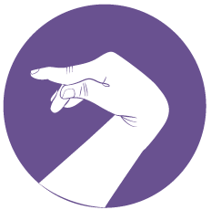 The wrist is usually flexed, and turned palm down (“pronation”).
It is difficult, and sometimes even impossible for the patient to bring the wrist into extension because of the hypertonic (spastic) flexor muscles. But it can be done passively, i.e. if one takes the wrist and brings it gently but firmly into extension.
Like in the elbow, clinical examination helps to distinguish between spasticity, and muscle contracture. If it is contracted, the wrist is permanently flexed, even at rest, and cannot be brought into extension passively. Because of this flexed position, it may be very difficult to move and use the fingers, even if they are otherwise normal.(picto 3: caroline 9) Correction of the deformity (physical therapy, splint, botulinum toxin, surgery) improves the function of the hand a great deal.

The “pronation” and the “ulnar deviation” deformities are frequent in spastic children.
With the arm resting along the body and the elbow flexed at a right angle (90°), pronation is the position of the hand palm down, as opposed to supination, which is the position palm up.
Ulnar deviation is the position where the wrist is neither flexed nor extended, but bent in the direction of the little finger, as opposed to radial deviation, where it is bent in the direction of the thumb. This deformity occurs less often in spastic adults.
Fingers
The position of the fingers is very variable.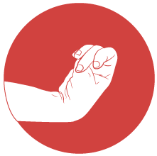
They can be closed, making a tight fist around the thumb, which is clenched into the palm. This position renders the hand useless. If there is only spasticity and no contracture, the fingers can usely be progressively brought into passive extension with firm but gentle traction. But the deformity soon recurs within a few minutes. When severe, this tight position may generate hygiene problems: unpleasant sweating, maceration, and even skin ulceration.
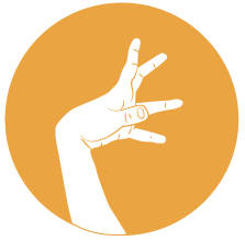
The fingers can also assume an extended and even hyperextended position. When the most proximal joints (metacarpo-phalangeal: MP) and the most distal joints (distal inter-phalangeal: DIP) are in a flexed position, and the middle joints (proximal inter-phalangeal : PIP) are in an hyperextended position, this is referred to as a “swan neck” deformity, due to an imbalance between two different muscle groups (extrinsic and intrinsic muscles).
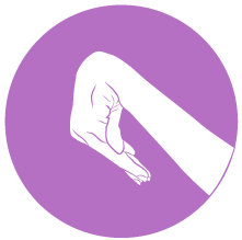
The function of the hand depends on finger deformities, but it is very much influenced as well by the position of the wrist. If the wrist is deformed in hyperflexion, it is mechanically impossible for the fingers to make a fist; even though they are otherwise able to move .
In a number of patients, correcting the position of the wrist improves drastically the capacity of the fingers to seize objects. This can be demonstrated by fitting the patient with a splint that immobilizes the wrist only.
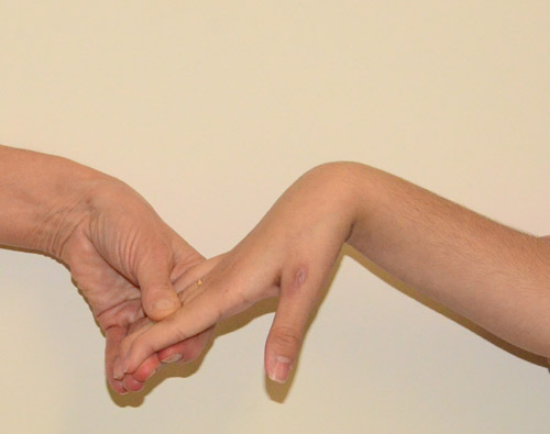
If the finger flexors (muscles which flex the fingers) are contracted, the fingers cannot be opened unless the wrist is brought in flexion.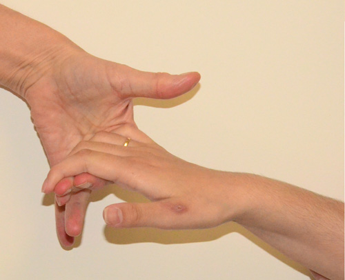
Thumb
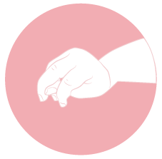
The thumb is often tight along the index finger, or even clenched into the palm because of spasticity of two muscle groups: the intrinsic muscles (adductor, short flexor, opponent) which cause the web space to be closely tight, and the extrinsic muscle (thumb long flexor) which causes the tip of the finger to flex.
This prevents opening of the thumb, even in cases where the muscles extending the thumb are active, and is a good example of the unbalance between the flexors and adductors, spastic and too strong, and the extensors, too weak to fight this hyperactivity. In such cases, not only is pinch between the thumb and the index finger impossible, but the thumb also gets in the way during grasping, preventing the use of the hand.
Children
Specific problems
Spasticity in children is mostly due to cerebral palsy. It occurs at birth or during the first two years of life. Once established, it is non progressive. It may impair one limb (arm or leg: monoplegia), two limbs (one side of the body: hemiplegia), or all four limbs (quadriplegia).
Children learn very early on to live with it and to adapt naturally to the deficit. Hemiplegic children develop amazing performances with the contra-lateral normal arm. They also naturally tend to ignore the impaired arm, despite functional possibilities. and may end up “forgetting “ it unless they are encouraged to use it. Occupational therapy plays an outstanding role in this education.This is not true of the spastic lower limb (leg) which is essential for walking.
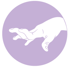 Therefore the upper limb must be managed early with physical and occupational therapy, in order to develop its potential and fully use its capacity, even though limited. Some of the specific problems in children affecting the upper limb include hyperlaxity of the finger joints (“swan neck” ) and the thumb, Abnormal movements
Abnormal movements, also referred to as “movement disorders”, occur involontarily and out of the patient’s control. They can occur at rest or during activity. In spastic patients, the most frequent abnormal movements are:
-dystonia, characterized by sustained muscle contractions cause twisting and repetitive movements or abnormal postures. Dystonia is often initiated or worsened by voluntary movements, and seems due to a lack of motor coordination (lack of reciprocal inhibition of agonist / antagonist muscles). In the upper limb it often produces abnormal postures in ulnar deviation of the wrist, and in finger extension when the wrist is flexed. Severe dystonia is a contra-indication to surgery.
- chorea is made of brief, semi-directed, irregular movements that are not repetitive or rhythmic, but appear to flow from one muscle to the next ('dance-like' movements).
- athetosis is characterized by slow, involuntary, convoluted, writhing movements of the fingers, hands, toes, and feet and in some cases, arms, legs, neck and tongue. It can be quite permanent, ceasing only during sleep. such as dystoniadystonia, characterized by sustained muscle contractions cause twisting and repetitive movements or abnormal postures. Dystonia is often initiated or worsened by voluntary movements, and seems due to a lack of motor coordination (lack of reciprocal inhibition of agonist / antagonist muscles). In the upper limb it often produces abnormal postures in ulnar deviation of the wrist, and in finger extension when the wrist is flexed. Severe dystonia is a contra-indication to surgery.. In selected cases, surgery can be very effective in restoring a good function, provided it is performed early, and followed by careful rehabilitation therapy
Timing of surgery
Spastic children usually require long term specific medical and paramedical care. Surgery may be necessary very early on if a deformity (i.e. severe elbow or wrist contracture) increases despite adequate rehabilitative treatment (physiotherapy and splinting).
In cases where the goal of surgery is to improve function, it can be performed as early as 6 years of age. If functional surgery is delayed until adolescence, by then the child has learnt other ways of prehension depending on the contralateral healthy limb, and when more normal patterns are restored through surgery, the older child may experience important difficulties in reprogramming his/her brain in order to achieve these “new“ functions.
Adult
Specific problems
The function of the spastic upper limb is extremely variable. Some patients use it almost normally, others do not use it at all (even if it has the capacity of moving). Many stroke patients have very little movement and a lack of sensation in the impaired limb, and they tend to “neglect” it. In these “non functioning” arms and hands, surgery has little to offer in terms of function. But it can be useful for improving severe deformities, such as elbow flexion, wrist flexion, finger flexion, and/ or thumb in palm. These deformities, which may due to spasticity alone, or associated with muscle or joint contractures, often affect adversely nursing, hygiene and cosmesis. They can most often be improved by surgery, with a real positive impact on the patient’s comfort, and family assistance .
Timing of surgery
Surgery can usually be programmed when there is no more progress in the motor / sensory recovery of the limb. In spasticity resulting from head injury (cerebrolesion), some severe deformities can develop very early on and keep worsening despite appropriate rehabilitative treatment. Such circumstances may require early surgical release.
Other problems
Spasticity may be associated with other deficits which impair function even more, such as abnormal movements (athetosisAthetosis is characterized by slow, involuntary, convoluted, writhing movements of the fingers, hands, toes, and feet and in some cases, arms, legs, neck and tongue. It can be quite permanent, ceasing only during sleep., chorea), vision deficiency, difficulties in walking and in body balance. Some patients will require crutches, walking aids or a wheelchair, which can limit even more the use of the hand.
Other problems associated with spasticity may include speach difficulty, hearing deficiencies, cognitive problems, emotional instability…
All of these problems must be taken into account when organizing the care of the individual person with spasticity.


